Development of photo-receptor protein based artificial receptive field and its application to image processing
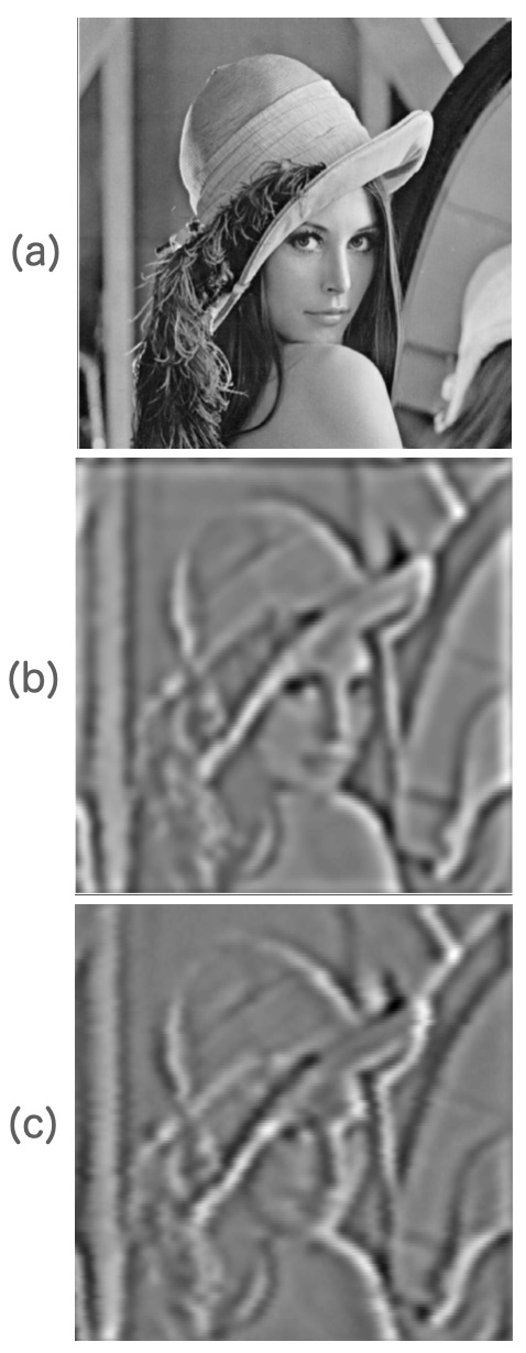
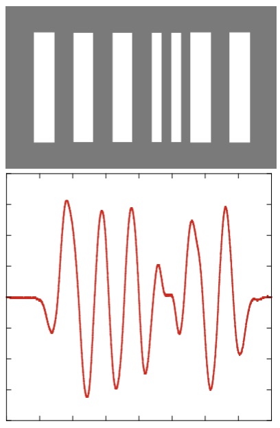
Visual information is first processed in retinal ganglion cell (RGC) transmitted to lateral geniculate nucleus (LGN) of the brain and then to primary visual cortex in the cerebrum. These visual cells have limited fields of view called receptive field which is arranged into the "centre" and the "surround", or the excitatory and the inhibitory region, each region responding oppositely to light.As shown in Fig.1, The shapes of receptive fields vary significantly depending on the region in which the neuron exists. Neurons in RGC and LGN have a coaxial-shaped receptive field structure which is modeld by DOG function. Simple cells have spatially oriented receptive fields with alternating oval. Bacteriorhodopsin, a visual function protein, shows differential responce with a polarization reversal and we interpret this response as an equivalent for excitatory/inhibitory response of receptive filed. Employing this particular feature we are aiming to implement receptive field type image processing device. Figure 2 shows the structure of the DOG filter and Gabor filter using bacteriorhodopsin. Figure 3 show the result of DOG filtering: (a) input image, (b) digital simularion, and (c) scanned by bR-DOG filter. Figure 4 shows an example of defect detection using the bR-Gabor filter. We are able to detect small differences of pitch just by scanning projected images.
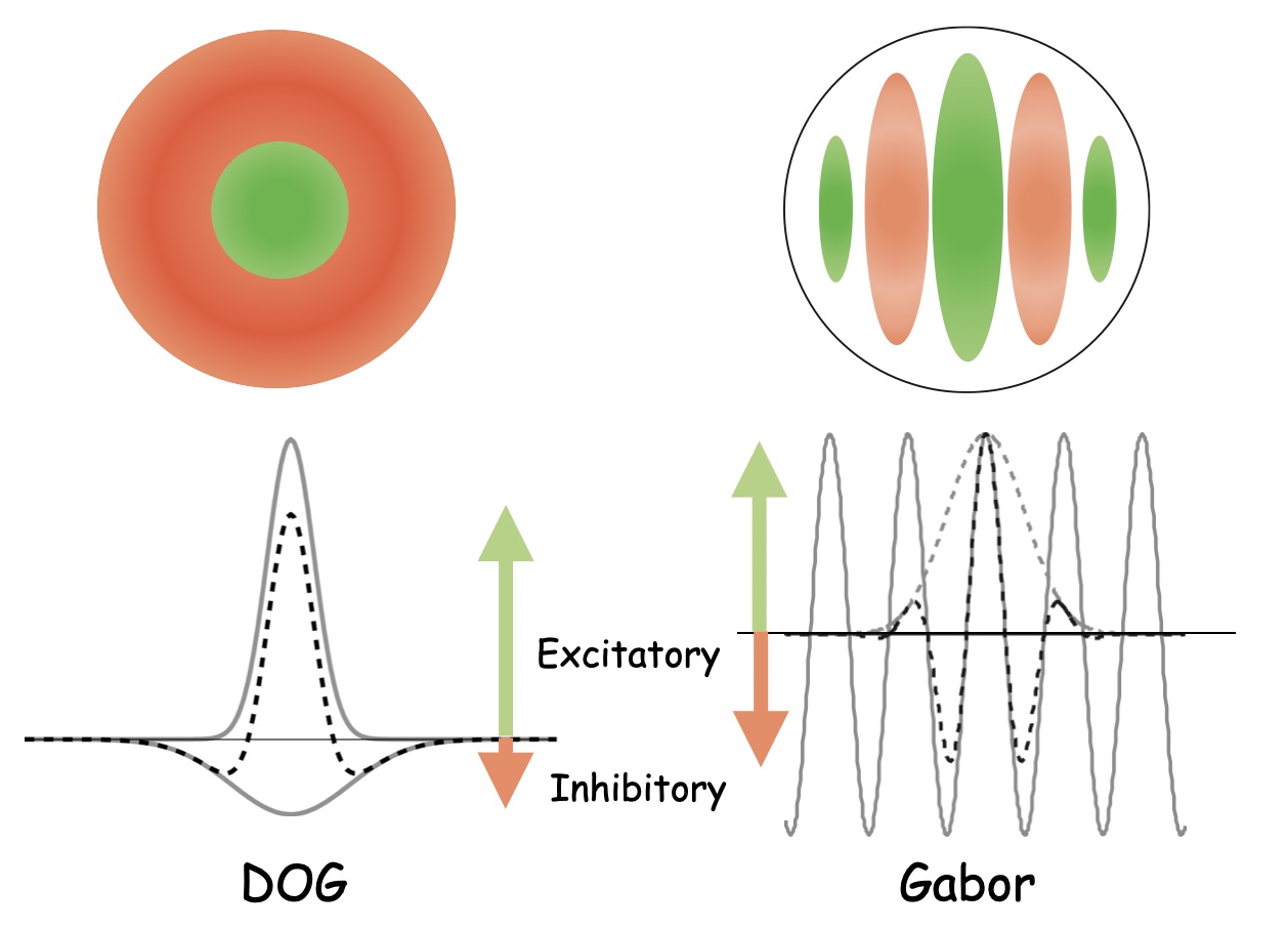
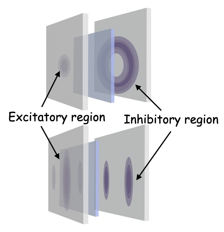
Fig.1 Fig.2